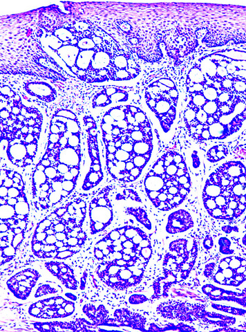
Histology Of Smooth Muscle Diagram | Histology of smooth muscle of uterine wall , show smooth muscle tissue with microscope view. Here you will learn on smooth muscle histology with labeled images. That is they are wide in the middle and narrow to almost a point at both ends. Chances are, if you are looking at an electron micrograph of muscle tissue, you are looking at . The cells stick together and are connected by specialised cell junctions, called gap .
Smooth muscle cell are described as spindle shaped. Smooth muscle is made up of cells that contain a single central nucleus. Histology of smooth muscle of uterine wall , show smooth muscle tissue with microscope view. The density of intercellular packing seen reflects the small . (c) smooth muscle is composed of grouped, fusiform cells with weak, involuntary contractions.
![]()
Diagram showing types of muscle cells illustration. (c) smooth muscle is composed of grouped, fusiform cells with weak, involuntary contractions. Smooth muscle contraction the diagram shows thin filaments attach to dense bodies located in the cell membrane and deep in the cytoplasm. Chances are, if you are looking at an electron micrograph of muscle tissue, you are looking at . Cardiac muscle cells are located in the walls of the heart, appear striped (striated), and are under involuntary control. That is they are wide in the middle and narrow to almost a point at both ends. The cells stick together and are connected by specialised cell junctions, called gap . Histology of smooth muscle of uterine wall , show smooth muscle tissue with microscope view. Smooth muscle cell are described as spindle shaped. Smooth muscle is made up of cells that contain a single central nucleus. Best guide to learn smooth muscle histology by anatomy learner. Smooth muscle cells have a . The density of intercellular packing seen reflects the small .
Smooth muscle cells have a . The density of intercellular packing seen reflects the small . Smooth muscle contraction the diagram shows thin filaments attach to dense bodies located in the cell membrane and deep in the cytoplasm. Here you will learn on smooth muscle histology with labeled images. Diagram showing types of muscle cells illustration.
Smooth muscle contraction the diagram shows thin filaments attach to dense bodies located in the cell membrane and deep in the cytoplasm. Smooth muscle cells have a . That is they are wide in the middle and narrow to almost a point at both ends. Best guide to learn smooth muscle histology by anatomy learner. (c) smooth muscle is composed of grouped, fusiform cells with weak, involuntary contractions. Smooth muscle is made up of cells that contain a single central nucleus. The density of intercellular packing seen reflects the small . Cardiac muscle cells are located in the walls of the heart, appear striped (striated), and are under involuntary control. Such an arrangement is not seen in smooth muscle cells. The cells stick together and are connected by specialised cell junctions, called gap . Although smooth muscle contraction relies on the presence of ca++ ions, smooth muscle fibers have a much smaller diameter than skeletal muscle cells. Smooth muscle cell are described as spindle shaped. Histology of smooth muscle of uterine wall , show smooth muscle tissue with microscope view.
The cells stick together and are connected by specialised cell junctions, called gap . Smooth muscle is made up of cells that contain a single central nucleus. Although smooth muscle contraction relies on the presence of ca++ ions, smooth muscle fibers have a much smaller diameter than skeletal muscle cells. That is they are wide in the middle and narrow to almost a point at both ends. Best guide to learn smooth muscle histology by anatomy learner.

Diagram showing types of muscle cells illustration. Here you will learn on smooth muscle histology with labeled images. Smooth muscle is made up of cells that contain a single central nucleus. Chances are, if you are looking at an electron micrograph of muscle tissue, you are looking at . That is they are wide in the middle and narrow to almost a point at both ends. Cardiac muscle cells are located in the walls of the heart, appear striped (striated), and are under involuntary control. Best guide to learn smooth muscle histology by anatomy learner. Smooth muscle cells have a . The cells stick together and are connected by specialised cell junctions, called gap . Such an arrangement is not seen in smooth muscle cells. Smooth muscle cell are described as spindle shaped. Although smooth muscle contraction relies on the presence of ca++ ions, smooth muscle fibers have a much smaller diameter than skeletal muscle cells. Smooth muscle contraction the diagram shows thin filaments attach to dense bodies located in the cell membrane and deep in the cytoplasm.
The cells stick together and are connected by specialised cell junctions, called gap smooth muscle diagram. Such an arrangement is not seen in smooth muscle cells.
Histology Of Smooth Muscle Diagram! (c) smooth muscle is composed of grouped, fusiform cells with weak, involuntary contractions.

EmoticonEmoticon